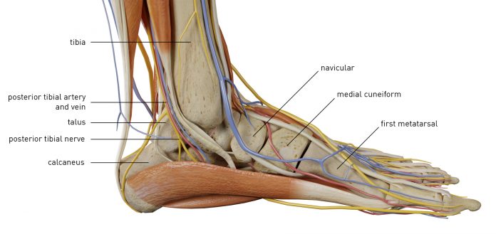
The ankle joint combines the fibula and tibia with the adrenal ankle anatomy gland-talus and foot bone.
The overgrown part of the bone enters the hole between the lower ankle anatomy of the fibula and tibia, near the junction of the formation of the ankle anatomy.
It is customary to highlight:
– Inner ankle – is the lower edge of the tibia;
– The outer the ankle is the edge of the fibula;
– The lower area of the tibia.
The ankle has a gap that forms on the inner surface of the upper side of the talus and hyaline cartilage.
The anatomy of the ankle anatomy is quite complex; in its area, muscles, bones, and ligaments are connected. Thanks to the ankle, a person can maintain balance and move in the usual ankle anatomy way. The muscular framework allows the bones to withstand significant loads and also protects the musculoskeletal system from injury. The surrounding blood vessels provide nutrition to the entire ligament of the ankle anatomy.
The anatomy is not simple, so it often causes damage due to heavy loads. To avoid injuries, complications, and inflammatory processes, you need to understand the structure of the ankle.
Foot anatomy
The calcaneus can be safely considered the most massive of all those located in the foot area. The anatomy of the calcaneus is unique; its primary function is a kind of springboard during movement. It is massive and has high endurance, but can be easily damaged under the influence of many factors. What pathologies in its area can be formed?
The calcaneus connects to other bones. It is located directly under the talus, which is connected to the heel by a short calcaneal process. Behind it, a powerful tubercle, from which the lateral and medial processes extend along the sole. The second is connected to the tendon of the flexor of the toes. Next, the connection of the lateral process with a long plantar ligament is seen. The upper surface of the calcaneus is protected by the posterior talus articular surface associated with the rear calcaneal articular surface of the subtalar joint.
The skeleton of the foot consists of three sections: tarsus, metatarsus, and toes.
Tarsal bones
The posterior tarsal region is composed of the talus and the calcaneus; the anterior one is scaphoid, cuboid, and three sphenoids.
The talus is located between the distal [e.g., end, phalanx] (distal) – the end of a muscle or bone of a limb or the whole structure (phalanx, power) farthest from the body. The antonym is proximal …. click for details… The end of the bones of the lower leg and the calcaneus, being a kind of bone meniscus between the bones lower legs and bones of the foot.
The talus has a body and a head, between which there is a narrowed place – the neck.
The body on the upper surface has a joint surface – the talus block, which serves to articulate with the bones of the lower leg. On the front cover of the head, there is also a joint surface for articulation with the scaphoid. The inner and outer surfaces of the body are articular surfaces articulating with the ankles; on the lower surface, there is a deep groove separating the articular surfaces, which serve to say it with the calcaneus.
The calcaneus is the posterior lower tarsus. It has an elongated, flattened shape laterally and is the largest of all the bones of the foot. It distinguishes between the body and the well-palpable calcaneal protuberance protruding posteriorly. This bone has articular surfaces that serve to articulate from above with the talus, and in front with the cuboid bone. Inside the calcaneus, there is a ledge – the support of the talus.
The scaphoid is located at the inner edge of the foot.
It lies in front of the ram, behind the sphenoid, and inside of the cuboid bones. At the inner edge, it has a tuberosity of the scaphoid, facing downwards, which is well palpable under the skin and serves as an identification point for determining the height of the inner part of the longitudinal arch of the foot. This bone is convex anteriorly. It has articular surfaces articulating with adjacent bones.
The cuboid bone is located at the outer edge of the foot and articulates behind the heel, inside with the scaphoid and outer sphenoid, and in front with the fourth and fifth metatarsal bones. On its lower surface, there is a groove in which the tendon of the long peroneal muscle lies.
The sphenoid bones (medial, intermediate, and lateral) lie in front of the scaphoid, inside of the cuboid, behind the first three metatarsal bones and make up the anteroposterior tarsus.
Metatarsal bones
Each of the five metatarsal bones has a tubular shape. They distinguish between the base, body, and head. The body of any metatarsal bone resembles a trihedral prism in shape. The longest bone is the second, the shortest, and the thickest is the first.
On the bases of the metatarsal bones, there are articular surfaces that serve to articulate with the bones of the tarsus, as well as with adjacent metatarsal bones, and on the heads are articular surfaces for articulation with the proximal [eg.end, phalanx] – (proximal) – a point on a limb (edge of a bone or muscle) or an entire structure (phalanx, power), less distant from the body. Antonym – distal … click for details .. phalanges of fingers. The bones are located in different planes and form an arch in the transverse direction.
Finger bones
Toes consist of phalanges. As on the brush, the first toe has two phalanges, and the rest – three. There are proximal, middle, and distal phalanges. Their significant difference from the phalanges of the brush is that they are short, especially the distal phalanges.
On foot, as well as on the hand, there are sesamoid bones. Here they are much better expressed. Most often, they are found in the area of connection of the first and fifth metatarsal bones with proximal phalanges.
Sesamoid bones increase the transverse arching of the metatarsus in its anterior section.
Before talking about the structure of various departments of the foot, it should be mentioned that in this department of the leg, bones, ligamentous structures, and muscle elements interact organically.
Tarsal bones articulate with the tibia elements in the ankle joint.
This element on each side connects to the fibula and tibia.
In the lateral parts of the joint, there are bone outgrowths – ankles. The inner is the tibia, and the outer is the fibula. The articulation is:
In shape – blocky.
In terms of movement – biaxial.
Post-injury rehabilitation
Rehabilitation is an important step in the treatment of ankle sprain. Unfortunately, giving universal recommendations for severe trauma to this joint will be quite difficult.
Walking
With mild stretching, restoration of ankle mobility should begin with normal walking, excluding jumping and running at the initial stage of rehabilitation.
The walking pace should be moderate, you need to walk at least 5 km a day. But not right away – start with short walks of 2-3 km.
After a walk, you should do a contrasting water procedure: douse your feet in succession with a cold shower, hot, cold again. This will help restore blood microcirculation and accelerate venous outflow.
For a month, your “training” should stretch for at least 7-10 km. The pace should be slightly faster than moderate.
Climbing toes
The next stage – to walks we add a rise in socks with a change in the position of the ankle: socks inward, socks apart, socks in a neutral position.
We perform each movement slowly, until a strong burning sensation in the area of the feet and calf muscles. This step will take 2 weeks.
Bone frame
The ankle the joint is located at the junction of the tibia and tibia, as well as the talus. Immediately there is the talus-fibular ligament. All these bones form a cavity in which the joint is directly located. They also assume the main load during movement, since it is the ankle region that accounts for the entire mass of the human body.
Muscles, joints, and tendons connect all the bones of the ankle and foot. This gives the foot flexibility and provides good cushioning while walking. The structure of the ankle joint is unique.
Ligaments
The anatomy of the ankle, ligaments are presented below.
They are responsible for the normal functioning and movement of the joint, and they also hold bone elements in place. The most powerful is the deltoid ligament. It connects the talus, calcaneus, and scaphoid bones (feet) with the inner ankle.
A powerful formation is the ligamentous apparatus of the tibiofibular syndesmosis.
Holding the tibia together provides the interosseous ligament, this is a kind of continuation of the interosseous membrane. It goes into the lower back, which prevents the joint from turning too hard inward. Outward rotation does not allow the anterior lower tibial ligament.
Its location is between the calcaneofibular ligament located on the surface of the tibia and the outer ankle. Excessive rotation of the foot outwards prevents the transverse ligament, which is located under the tibia.
Functions
Thanks to the coordinated work of the muscles in the joint, movements in two planes are possible. In the frontal axis of the human ankle performs flexion and extension. The vertical plane, rotation is possible: inward and in a small volume outward.
– providing mobility of the body;
– uniform distribution of human weight throughout the foot;
– amortization of sudden movements;
– reduces shaking that occurs while walking, running;
– provides body stability;
– Gives smooth movements when walking on stairs;
– provides stability to the body when moving on an uneven surface.
Diagnostics
In such a complex element of the musculoskeletal system as the ankle, various pathological processes can occur. To detect a defect, visualize it, correctly make a reliable diagnosis, there are various diagnostic methods:
Roentgenography. The most economical and affordable way to research. .
However, the method is good economical, quick, no harmful effects on the tissue. So, You can detect the accumulation of blood and swelling in the joint bag, foreign bodies, visualize the ligaments.
Before, CT scan. With fractures, neoplasms, arthrosis, this technique is the most valuable in the diagnostic plan.
Before, Magnetic resonance imaging. So, As with the study of the knee joint, this procedure is better than any other will indicate the condition of the articular cartilage, ligaments, Achilles tendon. So, the technique is expensive but as informative as possible.
Before, Arthroscopy Minimally invasive, low-traumatic procedure, which includes the introduction of a chamber into the capsule. So, the doctor can examine the inside of the bag with his own eyes and determine the focus of the pathology.


